Hysteroscopic Surgery in Mumbai, India

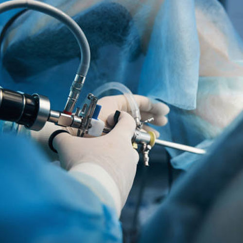
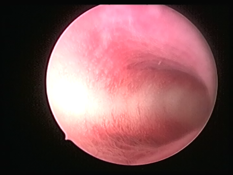
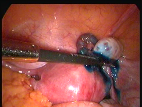
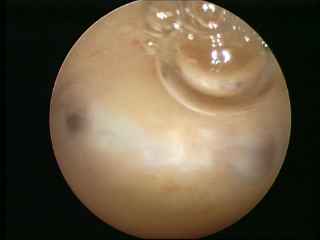
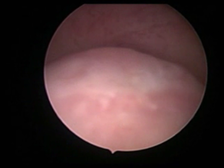
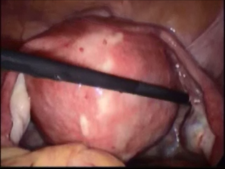
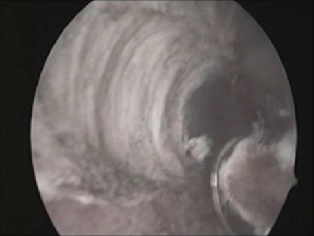
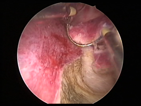
Hysteroscopy means peeping inside the uterine cavity with the help of a fine telescope varying between 2-4mm in diameter. Today a large number of surgeries can be performed via hysteroscopy due to advances in technology and instrumentation. These procedures do not involve any cut on the body and, hence, are virtually painless. The patient can be admitted on the day of the surgery and, if a laparoscopy is not needed, discharged in a few hours.

Hysteroscopy can be performed for the following indications :

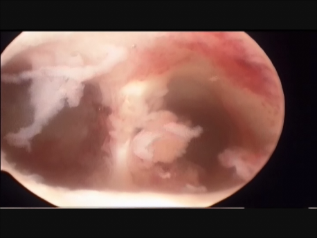
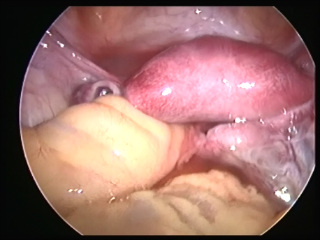
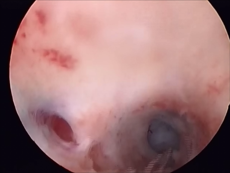
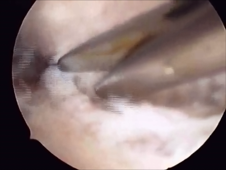
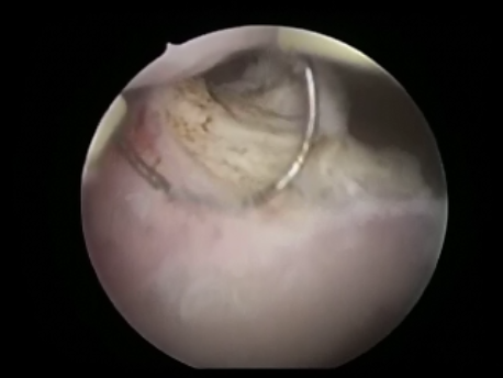
  • Infertility and recurrent loss (Subfertility): Infertility means the inability to conceive and recurrent pregnancy loss means loss of 3 or more (in practice, usually 2 or more) consecutive early pregnancy (less than 5 months). Uterine pathology such as abnormal shape (septum or the T shaped uterus), submucous fibroids or polyps, and intrauterine adhesions can cause subfertility. Tubal block due to spasm or mucus plugs can contribute to infertility and can be opened by hysteroscopic cannulation.
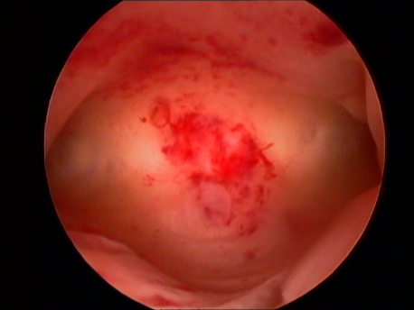
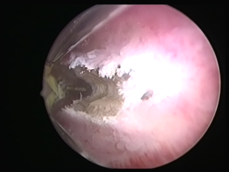
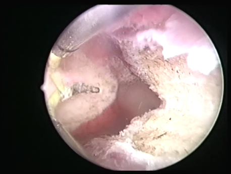
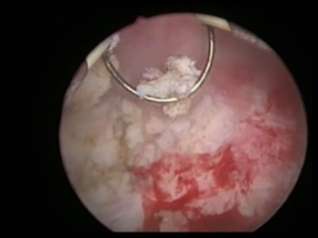
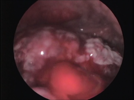
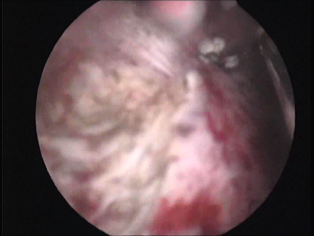
  • Abnormal uterine bleeding: Heavy, prolonged or irregular bleeding can be caused by submucous fibroids and polyps.
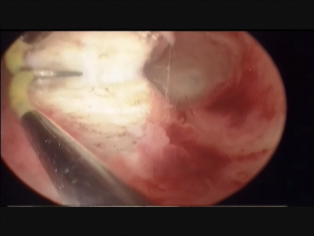
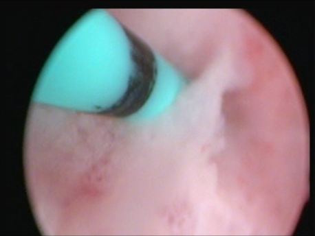
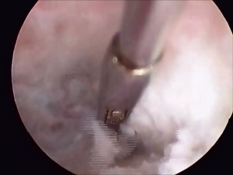
  • Intrauterine foreign bodies: Missing copper T embedded in the uterine wall or retained fetal bones or pregnancy following a miscarriage can contribute to irregular bleeding, infection or subfertility.
Hysteroscopic Surgery

The following surgeries can be performed by hysteroscopy :

  • Metroplasty for a uterine septum or a T-shaped uterus : A uterine septum is a congenital partition inside the uterine cavity which divides the cavity into two symmetrical halves. It may extend for varying lengths into the cavity, cervix and even the vagina. The septum can be cut by hysteroscopy by using an electrode which cuts the septum with an electric current or with fine hysteroscopic scissors. Since this surgery does not involve cutting the entire uterine wall, the uterus heals completely with minimum scarring and the woman can attempt a normal delivery during subsequent pregnancy. A normal uterine cavity is triangular in shape. When the side walls and the upper part of the uterus (fundus) converge, it is called as a T-shaped uterine cavity. This is associated with a high risk of pregnancy loss since the volume and the blood supply to the uterine cavity is reduced. The shape of the T-shaped uterus (triradiate or hypoplastic uterus) can be corrected by cutting the side walls and fundus with an electic current or hysteroscopic scissors. The pregnancy rates after these procedures are quite astonishing.
  • Hysteroscopic myomectomy & polypectomy : Sometimes fibroids (tumors of the uterine muscle or myometrium) can encroach on the uterine cavity. These are submucous fibroids and can lead to heavy menstrual bleeding, infertility and pregnancy loss. Similarly, localized overgrowth of the endometrium (inner lining of the uterine cavity) is called an endometrial polyp. This, too, can contribute to abnormal uterine bleeding and subfertility. These fibroids and polyps can be removed via hysteroscopy by cutting them into small pieces with an electric current (the resectoscope).
  • Adhesiolysis : Adhesions can form inside the uterine cavity causing the walls to stick to one another. This is due to previous infection (tuberculosis or pelvic inflammatory disease), previous uterine curettage or uterine surgery (myomectomy or hysterotomy or any surgery involving cuts on the uterus and entering the cavity). These adhesions lead to loss of menstruation (amenorrhoea), infertility and recurrent pregnancy loss. These adhesions can be cut via hysteroscopy using a fine scissors. If the cavity returns to normal, the woman can resume her menses spontaneously and can conceive (become pregnant) with IVF (test tube baby).
  • Removal of foreign bodies : A copper T device can get embedded in the uterine wall and prove difficult to remove manually. An incompletely aborted pregnancy can remain inside the uterus for years and cause abnormal uterine bleeding, pain and infertility. An ectopic pregnancy can implant away from the uterine cavity and into the cervix (mouth of the uterus) or the site of a cesarean section scar. These pathologies can be removed with the help of a hysteroscopic grasping forceps or resected out with an electrode.
  • Hysteroscopic tubal cannulation : The Fallopian tubes can be blocked because of tubal spasm or mucus plugs. The sperms ansd eggs cannot be transported across these blocked tubes. The tubal block can be opened by passing a guide wire into the Fallopian tube and a catheter over it via hysteroscopy. This is associated with high spontaneous pregnancy rates. However, pathologically damaged tubes (due to infection such as tuberculosis) cannot be opened by cannulation.
  • Transcervical resection of the endometrium (TCRE) : In women suffering from heavy menstrual bleeding with normal endometrium, the endometrium can be cut out (resected) with the means of an electrode and the basal layer burnt. This is an extremely effective procedure with prolonged relief from heavy menses.
  • Other procedures : Select fibroids can be removed by a hysteroscopic morcellator. This is a device which literally eats up the fibroid into small pieces with the help of a cutting end and a motor. The Fallopian tubes can be blocked (before IVF or for sterilization) with the help of a flexible spring loaded device (Essure) which is passed into the Fallopian tubes via the hysteroscope.

The advantages of hysteroscopic surgery include :

  • Absence of scar on the uterine muscle (hence better healing).
  • Absence of abdominal scar if laparoscopy is not indicated.
  • Minimal or no surgical pain.
  • Minimal anesthesia.
  • Day care procedure (admission on the day of surgery and discharge in a few hours in majority of cases).