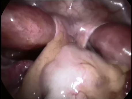
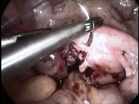
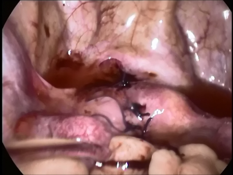
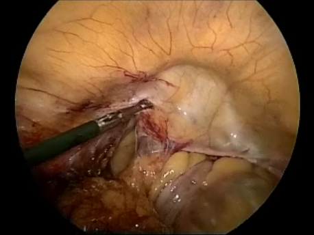
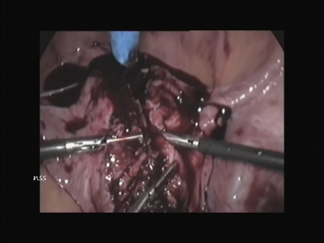
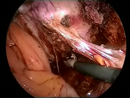
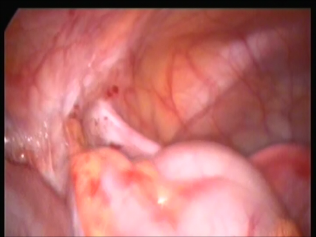
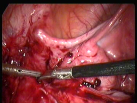
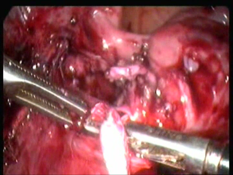
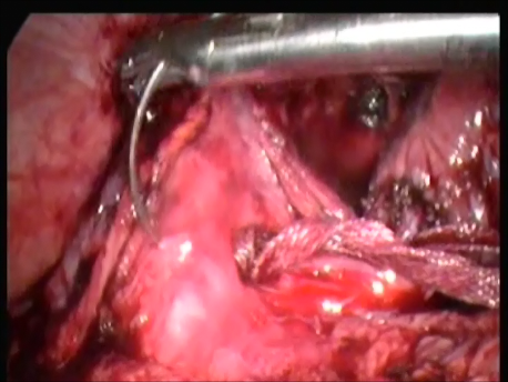
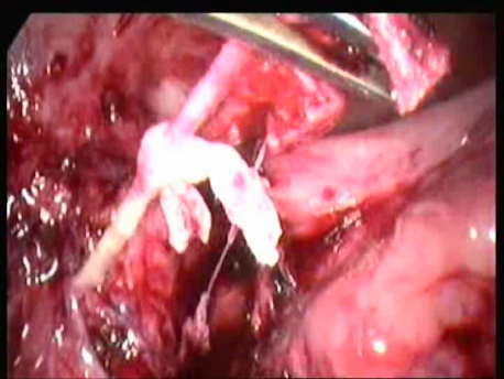
Laparoscopic release of entrapped genitofemoral nerve after shirodkar’s sling
Shirodkar’s sling is a surgery performed for treatment of pelvic organ prolapse. Occasionally, the genitofemoral nerve, a nerve passing along the side wall of the pelvis, may get entrapped in the tape used for the sling. This causes excruciating pain with difficulty in using that leg. We were referred a woman who had such a complication. Via laparoscopy, we released the nerve from the tape, cut the tape and placed a new tape at a different position to prevent recurrence of the prolapse. The woman recovered completely from her symptoms. This was the first ever reported case of its kind. It was presented at a national conference and is to be published a national journal shortly.
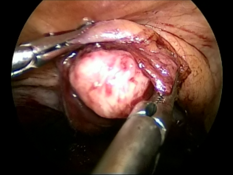
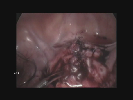
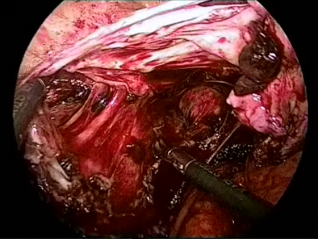
Laparoscopic removal of a parasitic fibroid invading the bladder
Occasionally, following hysterectomy or myomectomy, fibroid tissue (tumor of the uterine muscle) may implant inside the abdomen on the intestine, ovary, peritoneum or bladder and grow. We managed a rare case of a fibroid growing into the urinary bladder. We enucleated the fibroid completely from the bladder and the woman recovered completely from her urinary symptoms.
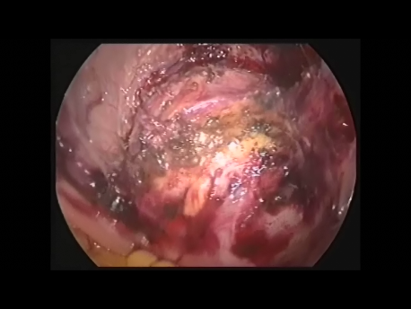
Laparoscopic excision of scar ectopic pregnancy
A scar ectopic pregnancy is a pregnancy which implants into the uterus at the site of a cesarean section scar. The pregnancy can invade the uterine wall and into the urinary bladder. Life threatening bleeding can occur. Hysterectomy is often necessary. We managed a case of scar ectopic pregnancy by laparoscopy. We first ligated the major blood vessels supplying the pelvis (internal iliac arteries), excised the ectopic pregnancy and repaired the cut in the uterus by absorbable sutures. The uterus could thus be saved with the hope for future fertility.
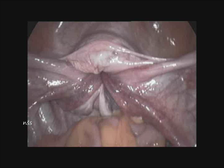
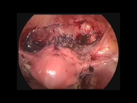
Laparoscopic unification of a bicornuate uterus
During development of the human body, the uterus consists of two halves which unify form a single uterus. Sometimes, the uterus may persist as two halves. This is called a bicornuate uterus. This is associated with an increased risk of miscarriage. Unification is a surgery whereby the two halves of the uterus are unified to create a single uterus with a single uterine cavity. This is done only for a woman with repeated miscarriages and NOT for a woman with infertility. We performed this surgery via laparoscopy whereby we incised the inner aspect (medial) of the two uteri till we entered each uterine cavity and sutured the two halves together with absorbable sutures. Repeat X-ray (hysterosalpingography) of the uterus or a hysteroscopy of the cases where we have done a unification have revealed normal uterine cavities.
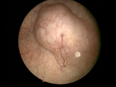
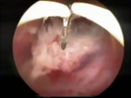
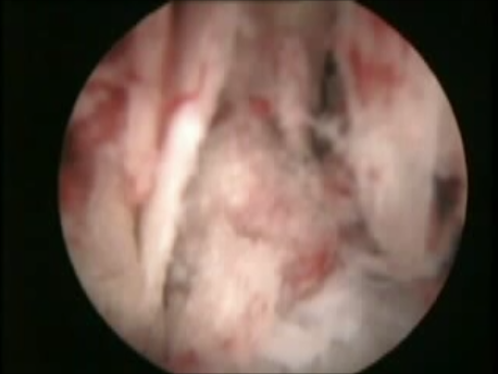
Hysteroscopic removal of fetal bones
Sometimes, a pregnancy at an advanced stage may undergo fetal demise (death). This pregnancy is usually spontaneously aborted by the uterus. Rarely, this pregnancy may persist. The soft tissues get absorbed and the fetal bones remain embedded inside the uterus. We managed such a woman who had stopped menstruating (amenorrhoea) and was being treated for infertility. Ultrasound showed calcification in the uterus and she had even been advised removal of the uterus! By using an operative hysteroscope, we excised all the bones in the uterine cavity. The woman started menstruating spontaneously and she had a spontaneous pregnancy and normal delivery.
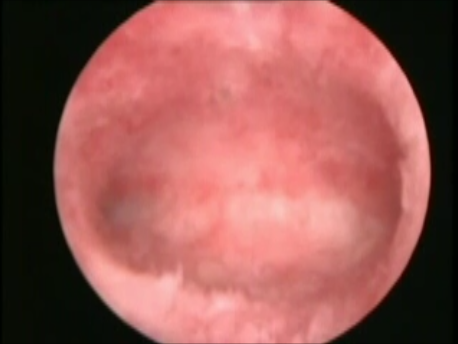
Hysteroscopic excision of a cervical ectopic pregnancy
Cervical ectopic pregnancy is a pregnancy which implants into the wall of the cervix. This is often mistaken for an incomplete miscarriage. The woman can experience massive uterine bleeding which may prove fatal. We treated a woman with a cervical ectopic pregnancy. We ligated the blood vessels supplying the cervix on either side ant then with the hysteroscope excised the pregnancy using and electrode. There was very little blood loss and the pregnancy could be removed completely.